|
|
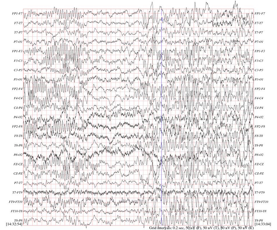
EEG (Electroencephalograph)
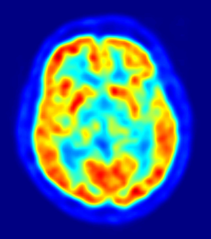
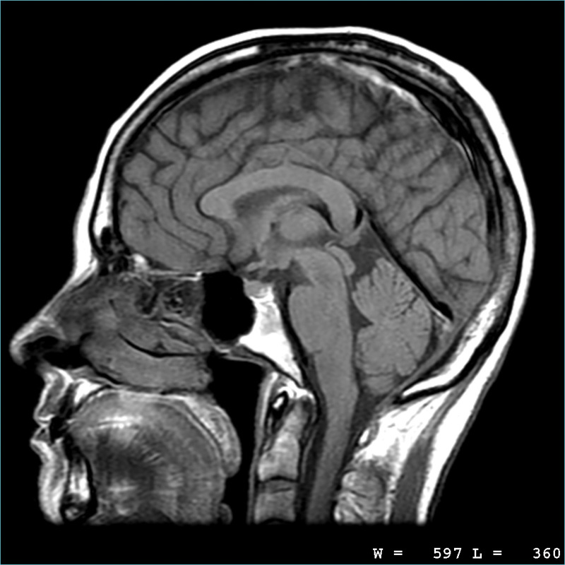
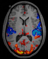
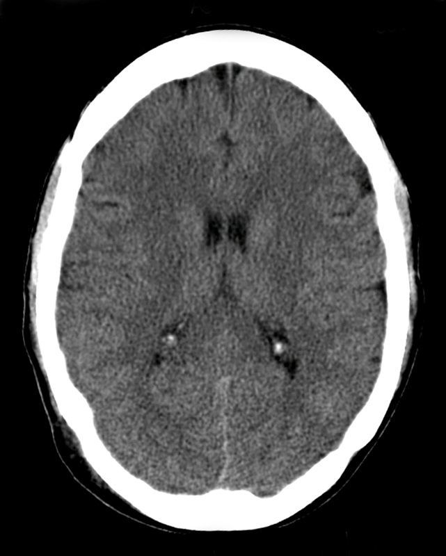
An amplified recording of the waves of electrical activity that sweep across the brain's surface. These waves are measured by electrodes placed on the scalp. The EEG is used to evaluate brain disorders. Most commonly it is used to show the type and location of the activity in the brain during seizures. It also is used to evaluate people who are having problems associated with brain function. PET (Positron Emission Tomography): A PET scan is a visual display of brain activity that detects where a radioactive form of glucose goes while the brain performs a given task. It is done to inspect the blood flow, oxygen intake or metabolism of tissues. Most commonly it is used to detect cancer, brain disorders and problems with the central nervous system. Unlike other imaging tests, such as CT or MRI scans, PET scans show abnormalities with tissues at the cellular level, MRI (Magnetic Resonance Imaging): A technique that uses magnetic fields and radio waves to produce computer-generated images of soft tissue. They scans show brain anatomy and is used to find problems such as tumors, bleeding, injury, blood vessel diseases, or infection. fMRI (Functional Magnetic Resonance Imaging): A technique for revealing bloodflow and, therefore, brain activity by comparing successive MRI scans. fMRI can be used to produce activation maps showing which parts of the brain are involved in a particular mental process. CT (Computed Topography) A series of X-ray photographs taken from different angles and combined by computer into a composite representation of a slice through the body. Also called CAT scan. |
Brain Imaging Techniques
EEG, PET, MRI, FMRI and cat